

Introduction
It grows slowly and very rarely spreads, but it can destroy surrounding tissue if left untreated. Early diagnosis and treatment lead to a complete cure in the vast majority of cases.
Symptoms
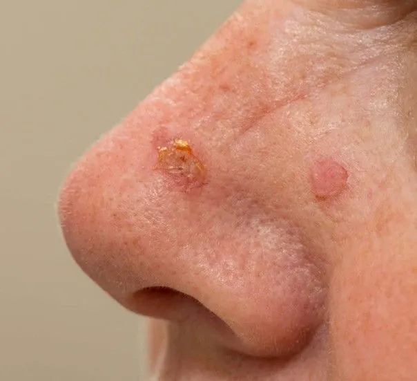
- Small pearly or translucent lesion
- Pinkish nodule with a shiny surface
- Crusted lesion that does not heal
- Chronic sore, sometimes ulcerated
- Visible small blood vessels on the surface (telangiectasias)
- Repeated minor bleeding
- Usually painless
- nose
- eyelids
- cheeks
- forehead
- ears
- balding scalp
Causes
- Chronic UV exposure
- Repeated sunburn
- Fair skin type (phototype)
- Older age
- Male sex
- Personal history of skin cancer
- Weakened immune system
Diagnosis
- Specialist dermatological examination
- Dermoscopy (characteristic features)
- Skin biopsy or diagnostic excision
- Staging investigations unnecessary in most cases
- Lesion persisting > 1 month
- Slow but steady growth
- Repeated bleeding
- Central ulceration
Treatments
- Standard surgical excision
- Mohs micrographic surgery (selected cases)
- Reconstruction if needed
- Specific topical treatments (superficial forms)
- Cryotherapy (limited forms)
- Radiotherapy in particular situations
- Photodynamic therapy (PDT) for certain superficial forms
- Selected dermatological lasers
- Medical LED to support healing
- complete removal of the tumour
Management
Specialist dermatology consultation
Histological confirmation
Individual choice of treatment
Early and complete treatment
Regular follow-up to detect recurrences or new lesions