

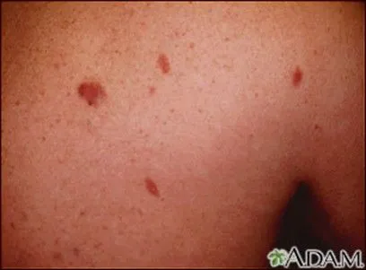
Introduction
Because they are rare, they can be difficult to diagnose, which is why specialist dermatological expertise and early multidisciplinary care are so important.
Symptoms
- Rapidly growing skin nodule or plaque
- Firm, infiltrated lesion, sometimes painful
- Unusual colour: purplish, dark red, skin-coloured
- Possible ulceration or bleeding
- Single or multiple lesions
- No improvement despite standard treatment
- Possible associated swollen lymph nodes
Causes
- Weakened immune system (organ transplant, immunosuppressive treatment)
- Older age
- Chronic sun exposure (for certain types)
- Cancer-causing viral infections (e.g. polyomavirus)
- Particular genetic background
- Merkel cell carcinoma (aggressive, fast-growing neuroendocrine cancer)
- Skin sarcomas (angiosarcoma, dermatofibrosarcoma, etc.)
- Skin adnexal tumours (sweat glands, sebaceous glands)
- Cutaneous lymphomas (primary T-cell or B-cell lymphomas of the skin)
- Skin metastases from an internal cancer
Diagnosis
- Expert dermatological examination
- Dermoscopy (helpful but not specific)
- Deep skin biopsy (essential)
- Histological and immunohistochemical analyses
- Routine staging investigations
- Review at a specialist multidisciplinary team meeting
Treatments
- Wide surgical excision (when possible)
- Adjuvant or primary radiotherapy
- Specialist systemic treatments (depending on the tumour type)
- Personalised combined protocols
- Supportive dermatological care
- Lasers and local techniques only in very selected cases
- Medical LED possible to support healing, never as a cure
Management
Rapid referral to a specialist dermatologist
Accurate histological diagnosis
Coordination with oncology, surgery and radiotherapy
Close, long-term follow-up
Clear information and support for the patient