

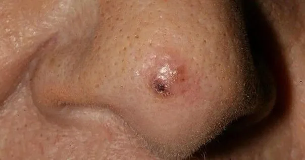
Introduction
Early detection and treatment determine the outlook.
Symptoms
- Firm lesion, sometimes painful
- Red, thickened patch or nodule
- Persistent crusts, cracks
- Ulcer or sore that does not heal
- Bleeding at the slightest knock
- Gradual, sometimes rapid growth
- Possible swollen regional lymph nodes in advanced forms
- face
- balding scalp
- lips (especially the lower lip)
- ears
- backs of the hands
- forearms
Causes
- Cumulative sun exposure
- Pre-existing actinic keratoses
- Fair skin type (phototype)
- Older age
- Male sex
- Weakened immune system (organ transplant, immunosuppressive treatment)
- Chronic scars, long-standing wounds
- Cancer-causing viral infections (HPV, in certain settings)
Diagnosis
- Specialist dermatological examination
- Dermoscopy to guide suspicion
- Skin biopsy or excision for histological confirmation
- Assessment of depth and high-risk features
- Staging investigations if risk criteria are present (invasive forms)
- Large size
- Considerable tumour thickness
- High-risk site (lip, ear)
- Pain, local paralysis
- Weakened immune system
Treatments
- Complete surgical excision with appropriate margins
- Mohs micrographic surgery
- Adjuvant or primary radiotherapy (specific situations)
- Topical treatments only for very limited superficial lesions
- Lymph node dissection if regional nodes are involved
- Photodynamic therapy (early superficial forms)
- Selected dermatological lasers
- Medical LED to support healing and reduce inflammation
- complete and rapid removal
Management
Urgent specialist dermatology consultation
Histological confirmation
Individualised treatment strategy
Multidisciplinary coordination if needed
Long-term regular follow-up to detect recurrence and metastasis