

Introduction
Early detection is crucial: when diagnosed at an early stage, melanoma can be cured in the vast majority of cases.
Symptoms
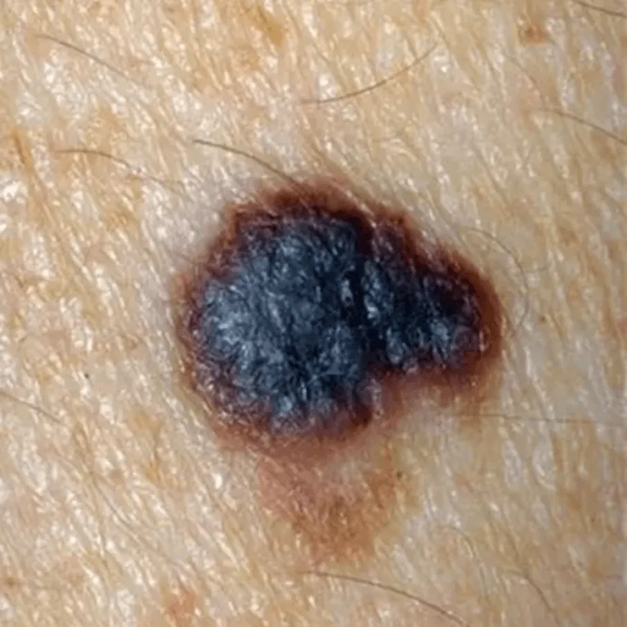
- A – Asymmetry: irregular shape
- B – Border: poorly defined, jagged
- C – Colour: uneven (brown, black, blue, red)
- D – Diameter: > 6 mm (but can be smaller)
- E – Evolution: rapid change
- Spontaneous bleeding
- Itching or pain
- Rapid growth
- Non-pigmented melanoma (rare but misleading)
Causes
- Intense sun exposure and sunburn, especially in childhood
- Fair skin type (phototype)
- Personal or family history of melanoma
- Large number of naevi
- Atypical naevi
- Weakened immune system
- Use of sunbeds
Diagnosis
- Full dermatological skin examination
- Dermoscopy (specific criteria for malignancy)
- Photographs and comparison over time
- Complete surgical excision of the suspicious lesion (histological examination essential)
- Assessment of tumour thickness (Breslow depth)
- Staging investigations depending on the stage
- Multidisciplinary discussion if necessary
Treatments
- Complete surgical excision with appropriate margins
- Wider re-excision
- Sentinel lymph node biopsy
- Specialist systemic treatments (advanced forms)
- Coordinated oncology follow-up
- stage of the melanoma
- strictly contraindicated
Management
Urgent specialist dermatology consultation
Rapid, documented diagnosis
Excision under optimal conditions
Coordination with oncology if necessary
Long-term, structured follow-up