

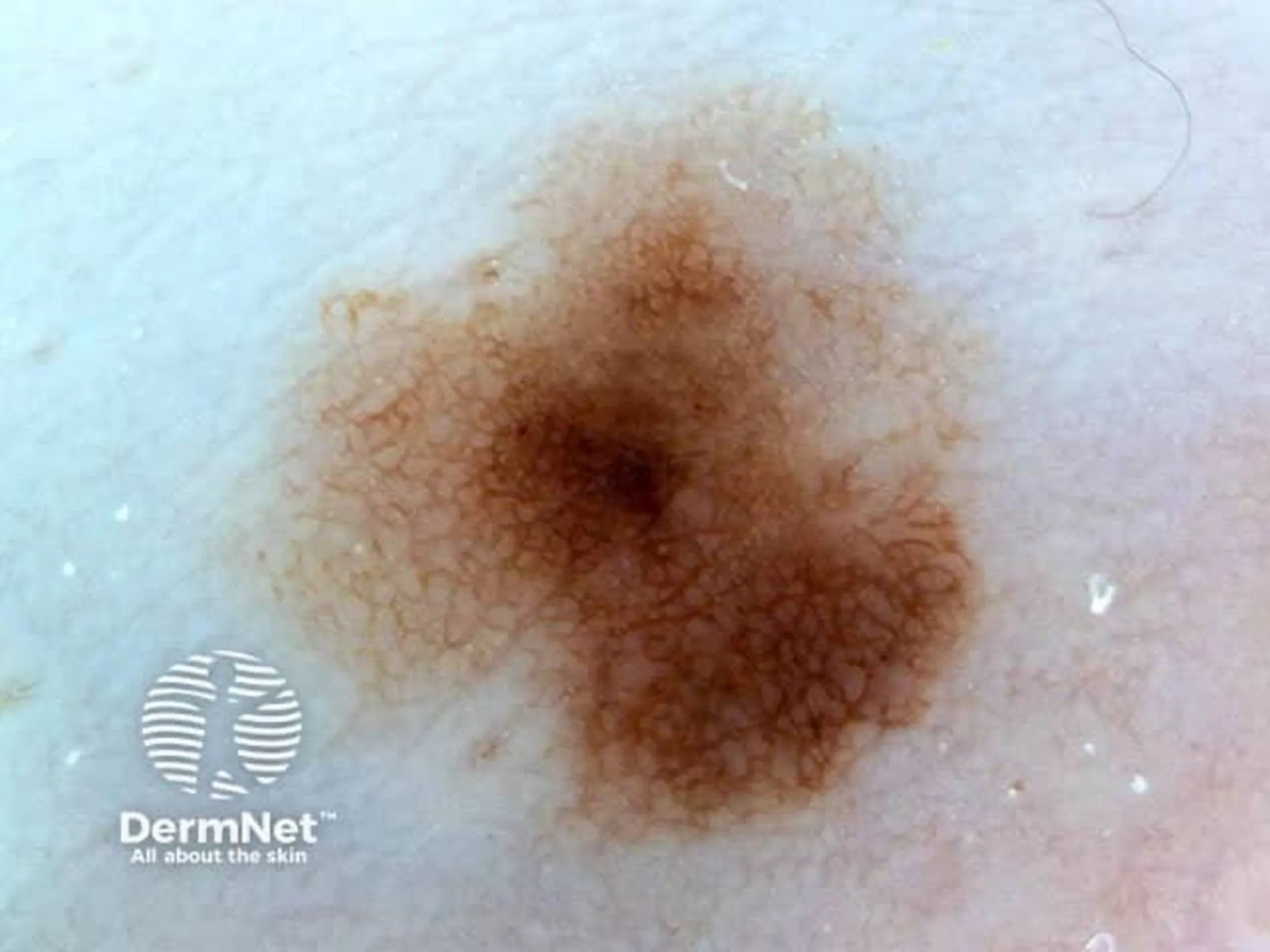
Introduction
Benign skin tumours are common skin lesions that are not cancerous. They account for a very large share of dermatology consultations, often for a diagnostic opinion, cosmetic concerns or worry about a recent change. The main challenge is to distinguish them with certainty from malignant lesions, in order to reassure the patient or arrange appropriate treatment if necessary.
Symptoms
- Pigmented or non-pigmented lesions
- Raised or flat (flat, dome-shaped, on a stalk)
- Variable colour: skin-coloured, brown, black, red
- Stable size or slow growth
- Usually painless
- No spontaneous bleeding (except after injury)
- Possible cosmetic or mechanical discomfort (rubbing)
Causes
- Genetic factors (naevi, skin tags)
- Skin ageing
- Chronic sun exposure
- Hormonal factors
- Benign proliferation of
- melanocytes keratinocytes vascular cells fatty tissue
- melanocytes
- keratinocytes
- vascular cells
- fatty tissue
Diagnosis
- Dermatological clinical examination
- Dermoscopy to analyse the structure of the lesion
- Comparison with the criteria for benign lesions
- Follow-up photographs if necessary
- Biopsy or diagnostic excision if in doubt
Treatments
- Cosmetic concerns
- Functional discomfort (rubbing, irritation)
- Repeated bleeding
- Diagnostic doubt
- At the patient’s request, after being informed
- Simple monitoring
- Local surgical excision
- Electrocoagulation
- Dermatological laser, depending on the indication
- Cryotherapy for certain lesions
- not always necessary
Management
Specialist dermatology consultation
An accurate, clearly explained diagnosis
Shared decision between monitoring and treatment
Procedures carried out under safe medical conditions
Follow-up if necessary