

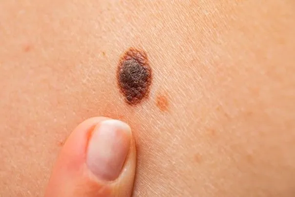
Introduction
Pigmented naevi, commonly known as moles, are very common skin lesions and are usually benign. They are a localised proliferation of melanocytes and may be present from birth or appear later in life. Dermatological monitoring is essential to detect any suspicious change.
Symptoms
- Pigmented spot or lesion, flat or slightly raised
- Variable colour: light brown, dark brown, sometimes black
- Size usually stable
- Regular borders in most cases
- Can occur anywhere on the body
- No pain or itching
Causes
- Genetic factors
- Localised accumulation of melanocytes
- Gradual appearance during childhood and adulthood
- Influence of sun exposure on the number of naevi
Diagnosis
- Clinical skin examination
- Assessment of size, colour and borders
- Dermoscopy, an essential tool for assessing the structure of the naevus
- Monitoring over time
Treatments
- Regular clinical monitoring
- Surgical excision if there is diagnostic doubt or the mole is bothersome
Management
A personalised dermatological assessment
Monitoring tailored to the patient’s profile
Clear information on warning signs
A medical decision if a suspicious change occurs